
Current Studies Examining 3D Printing for ACL Reconstruction
More than 150,000 Americans suffer from anterior cruciate ligament, or ACL, injuries each year, according to a recent report from American Orthopedic Society for Sports Medicine. The cost of those injuries? An estimated $500 million.
A Florida Tech research project, “3D Printed Biomimetic Bioglass-gradient Matrices for ACL Reconstruction,” funded by a $377,556 National Institutes of Health grant is exploring how to improve the clinical outcomes of ACL reconstruction. Vipuil Kishore, an associate professor in the chemical engineering program at Florida Tech, is leading the research.
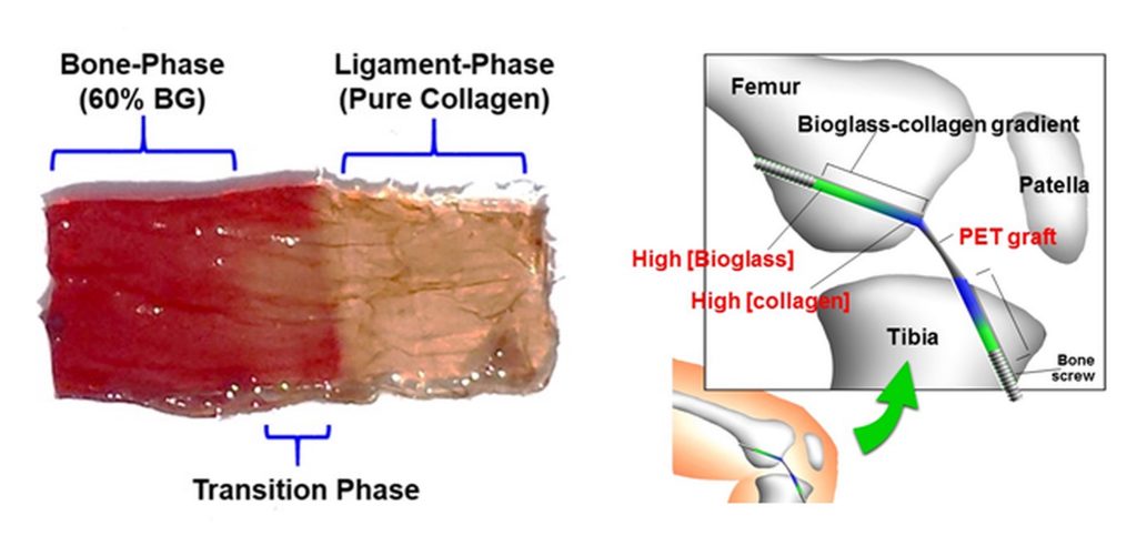
ACL injuries are highly common at the “enthesis region,” where the soft ligament meets the hard bone tissue. The goal of the research is to recreate the enthesis region at the bone-ligament interface using an innovative biomimetic approach that combines advanced technologies such as Raman spectroscopy and 3D printing. This project is attempting to use mixtures of collagen (ligament) and Bioglass (bone) as “bio-inks” to 3D print biomimetic-gradient scaffolds that simulate the native bone-ligament interface.
“A biomimetic approach entails generating scaffolds that mimic the native tissue as best you can, so you try to mimic the composition, structure and the mechanical aspects of the native tissue that you’re trying to replace,” Kishore said.
The ultimate goal of the research, and the reason why it is important, is to provide a new, potentially safer, solution to ACL injuries. Currently ACL surgeries are conducted primarily in two ways: with ligaments from elsewhere in the body or ligaments from cadavers. Both situations present significant risks: using another ligament could injure the body part it was taken from, and the body’s immune system could reject the cadaver tissue used.
“Several research groups across the United States and the world are working on devising new strategies so that we can make strides towards a common goal where one day we have an ACL solution that allows an athlete or an elderly person who has an ACL injury to return to a normal life after successful ACL reconstruction,” Kishore said.
In order to determine the composition of the bone-ligament interface, Kishore is using ACLs from rabbit cadavers to perform Raman spectroscopy, a laser-based, non-invasive technology that provides a structural fingerprint by which molecules can be identified. Raman data of the bone-ligament interface region can be converted to a stereolithography file, which provides a map for the 3D printer to print a biomimetic gradient using collagen and Bioglass inks called BioGIMs for “Bioglass gradient incorporated collagen matrices.”
In a design-build-validate strategy, Raman spectroscopy will then be used again to ensure the manufactured BioGIMs are an accurate replica of the natural bone-ligament interface.
Since cells have the ability to sense and respond to the surrounding environment, Kishore plans to test the ability of human stem cells to respond to the different biomimetic cues provided by the BioGIMs. The goal is to direct the cells to grow and mature as they would naturally in the body to enable regeneration of new, functional ACL enthesis tissue. Kishore has tested the stem cells on the printed collagen and Bioglass structures and has seen positive results.
“After testing the interaction with stems cells and the created gradient, future studies will entail integrating the printed gradients onto synthetic ACL grafts and assessing the functionality of the gradients in animal studies,” Kishore said. “There is still a lot of work to be done but development of new tissue engineering based strategies like the one we are working on in this research has significant potential to provide a viable clinical solution for implementation of artificial ligaments.”
###